Importance of Epicardial Adipose Tissue Localization using Cardiac Magnetic Resonance Imaging in patients with HFmrEF and HFpEF
In a study by Gijs van Woerden and Daan Westenbrink published in Clinical Cardiology, it was demonstrated that local-, rather than global Epicardial Adipose Tissue (EAT) is involved in the pathophysiology of patients with Heart Failure and Mid-Range or Preserved Ejection fraction (HFmrEF;HFpEF). This finding underscores the importance of quantifying EAT volume around the right ventricle, left ventricle and atria separately in addition to quantifying the total amount of EAT. Other members from Groningen Cardiology involved in this project included Dirk Jan van Veldhuisen, Thomas Gorter and Michiel Rienstra.
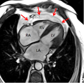
Epicardial Adipose Tissue (EAT) is becoming increasingly recognized as an important factor in the Heart Failure with Mid-Range and Preserved Ejection Fraction (HFmrEF; HFpEF) syndrome as it may exert negative effects on the underlying heart. However, it is unclear whether the adverse effects of EAT are due to a global-, or local mechanism. We precisely measured total EAT volume, as well as EAT volume surrounding the right ventricle, left ventricle and atria separately using cardiac magnetic resonance (CMR) imaging. We then related EAT to cardiac structure and function.
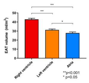
In 102 patients with HFmrEF and HFpEF, we found that the majority of EAT volume was located around the right ventricle. In addition, we showed that local EAT was associated with adverse cardiac remodeling and atrial fibrillation, and this association was independent of total EAT volume. These findings provide ground that local-, rather than global EAT volume is involved in the pathophysiology of HFmrEF and HFpEF. Therefore, precise localization of EAT is important and can easily be done using CMR imaging. Furthermore, these data give a greater understanding of the mechanism linking EAT to HFpEF/HFmrEF pathophysiology and hopefully lead to the development of new therapies for these HF patients.